T1 Mapping
CMR has the capability to characterise myocardial tissue using T1 and T2 mapping techniques. Quantitative T1 imaging, in particular, can be used to calculate the myocardial extracellular volume fraction (ECV), a measure of microscopic myocardial remodelling that has been associated with underlying diffuse fibrosis
Diffuse myocardial fibrosis is an underlying contributor to early diabetic cardiomyopathy, as it was documented by the association between myocardial diastolic dysfunction, post-contrast T1 values and metabolic disturbance.59 Recently, it was documented that non-contrast T1 mapping had high diagnostic accuracy for detecting cardiac AL amyloidosis, correlated well with markers of systolic and diastolic dysfunction and was potentially more sensitive for detecting early disease than LGE imaging; therefore, elevated myocardial T1 may represent a direct marker of cardiac amyloid load.60 In patients with SLE without cardiac symptoms, T1 mapping may detect subclinical myocardial involvement.61
Myocardial Inflammation
CMR is the ideal technique for the evaluation of inflammatory processes involving the heart. Myocarditis often has a subclinical course, which cannot be easily detected with standard inflammatory indices evaluated in the blood (erythrocyte sedimentation rate [ESR], C-reactive protein [CRP], etc.) and can lead, under special conditions, to dilated cardiomyopathy.62 During its early stages it can also remain undetected by the commonly used imaging techniques (echocardiography or nuclear imaging techniques), because these techniques are unable to distinguish slight tissue structure changes (oedema, cell infiltration), which can occur without associated changes in LVEF, the most often detected parameter by echocardiography. Instead, CMR can perform tissue characterisation, which makes it extremely valuable in myocarditis.
According to current experience, in myocardial inflammation due to infectious causes (i.e. viral myocarditis), a decrease in LVEF was not evident during the course of the disease, while an increase in cardiac troponin was found in only 20 % of cases.20 Additionally, myocardial biopsy is an invasive procedure and according to the American College of Cardiology (ACC)/American Heart Association (AHA) guidelines should be kept only for patients with unexplained new-onset heart failure <2 weeks in duration associated with a normal-sized or dilated left ventricle in addition to haemodynamic compromise, and cannot be used for screening or follow-up tool.62 Furthermore its diagnostic value is limited due to a number of reasons (sampling error, variation in observer expertise, etc.).61
CMR contributes to the diagnosis of myocarditis using three types of images: T2-weighted (T2W), early T1- weighted (T1W) images taken at one minute and delayed enhanced images (LGE) taken 15 minutes after the injection of contrast agent.
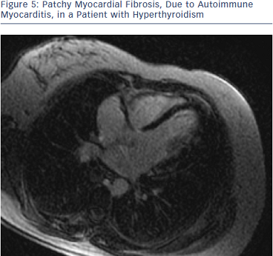
T2W is an indicator of tissue water content, which is increased in inflammation or necrosis, such as during myocardial infarction or myocarditis. However, it is not possible to differentiate between necrosis and inflammation only by the use of T2W images. To enhance the detection of pathology on CMR, images after early and delayed gadolinium injection should be obtained. Higher levels of early myocardial enhancement after gadolinium administration are due to increased membrane permeability or capillary blood flow. Membrane permeability is a major contributor as inflammation damages cell membranes through both T-cell perforin and B-cell antibody/ complement-mediated processes. The third parameter, which should be also evaluated, is the presence of contrast agent deposition in the delayed images (LGE images) (see Figure 5). Myocardial necrosis in the acute phase appears to play a major role in LGE formation, but also severe oedema could increase the gadolinium distribution and cause delayed enhanced areas. A combined CMR approach using T2W, early and late gadolinium enhancement, has a sensitivity of 76 %, a specificity of 95.5 % and a diagnostic accuracy of 85 % for the detection of myocardial inflammation.62,63
Evaluation of inflammation by CMR has already been applied in pheochromocytoma,64 diabetes mellitus,65 endocrine2 and rheumatic diseases,4 and allowed the detection of pathophysiology of cardiac lesions as well as cardiac disease acuity.
Limitations of Cardiac Magnetic Resources