
Intravascular ultrasound (IVUS) is the first catheter-based imaging modality that has been widely used in interventional c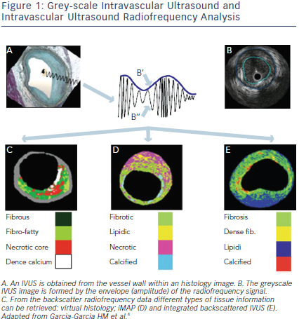 ardiology.1 It creates cross-sectional images of the vessel lumen and the arterial wall and provides valuable diagnostic information to angiography regarding lumen and vessel measurements and plaque morphology. This information helps clinical decision-making in ambiguous angiographic lesions. IVUS has also been used to evaluate new interventional devices and for atherosclerosis progression-regression trials. Recently, IVUS has been used to detect vulnerable plaques. In this review, we will focus on the potential clinical and research utility of IVUS-based imaging modalities for tissue characterisation.
Greyscale Ivus and Ivus Radiofrequency Analysis
IVUS uses a miniaturised piezoelectric transducer mounted on the tip of a catheter where it produces ultrasound signals. The imaging is based on the emission, attenuation and backscattering of ultrasonic waves that are converted to electrical signals and then processed as an image. Standard greyscale IVUS allows quantitative measurements of lumen and vessel and qualitative assessment of atherosclerotic plaque. Calcified tissues are highly echogenic and thus appear as bright echoes with acoustic shadowing. Regions of low echogenicity are usually labelled as “soft” plaque. Soft plaque has high lipid content or it may also be attributable to a necrotic zone within the plaque, an intramural haemorrhage or a thrombus. Fibrous plaques have an intermediate echogenicity between soft and calcific plaques.2 Recent IVUS studies have described attenuated plaque defined as hypoechoic plaque with deep ultraouns attenuation without calcification or very dense fibrous plaque.3 The grey-scale IVUS image is formed using the envelope (amplitude) of the radiofrequency (RF) signal. However, the frequency and power of the signal differ between tissues, regardless of similarities in the amplitude. Therefore, grey-scale IVUS has limited value for the accurate identification of specific plaque components. This limitation has been partially overcome by introduction of several IVUS-based post-processing methods. Analysis of IVUS radiofrequency backscatter enables a more detailed characterisation of plaque morphology or tissue characterisation and provides insight on the features of vulnerable plaque.4 Three different mathematical methods that have been applied to IVUS RF data analysis are virtual histology IVUS, Volcano Therapeutics (Rancho Cordova, CA, USA), iMAP-IVUS, Boston Scientific Corp (Santa Clara, CA, USA), Integrated Backscatter IVUS (YD, Nara, Japan) (See Figure 1, Table 1).