Scenario V – Stent Deployment and Edge Dissection

Add this Moment to your Passport

Learn from this moment and keep it forever.
FREE
Add To Passport

Preview

Summary

Scenario V – Stent Deployment and Edge Dissection Image titleImage title
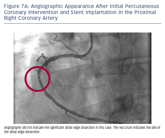
As an example of the utility of OCT and the superior resolution of this technology, it has become possible to diagnose potentially important problems after stent deployment; such as edge dissection, tissue prolapse or lack of lesion coverage. The significance of these findings remains to be demonstrated, although we have found it is not uncommon to find subtle examples of these problems after stent deployment that appear to be of little or no clinical consequence. However, when OCT imaging identifies a major problem after stent deployment, such as the case below, generally our practice is to address this issue.

Case 7
Age: 47
Gender: Male
History: Hypertension. Presented to an outside hospital and was ruled in for non-ST-segment myocardial infarction with a peak troponin I of 9.95 and an EKG showing inferior T-wave inversion.Image titleImage title

  • Cardiac catheterisation demonstrated the culprit lesion to be a thrombotic total occlusion of the right posterior descending artery (RPDA) branch, with an additional long 80–90 % lesion in the proximal RCA with a pre-PCI minimal CSA of 2.0 mm2 by OCT (not shown).
  • A single 3.0 x 38 mm stent was deployed across the proximal RCA lesion at 10 atmospheres.
  • Angiography suggested a well-deployed stent with no appreciable issues at the distal edge of the stent (see Figure 7A). OCT, however, showed a significant distal edge dissection (see Figure 7B).
  • Even though it was not seen on angiography, based on the OCT findings a decision was made to implant a second overlapping stent. A 3.0 x 23 mm stent was then deployed in the mid-RCA, overlapping with the first stent and covering the edge dissection (see Figure 7C).
  • Final OCT demonstrated complete coverage of the dissected region and good apposition of the stented segment (see Figure 7D).
  • OCT was also used to guide PCI of the culprit occlusion of the RPDA, which was completed uneventfully.

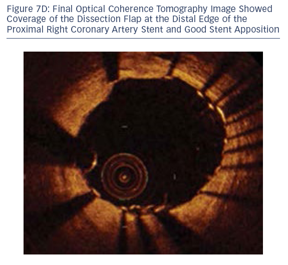
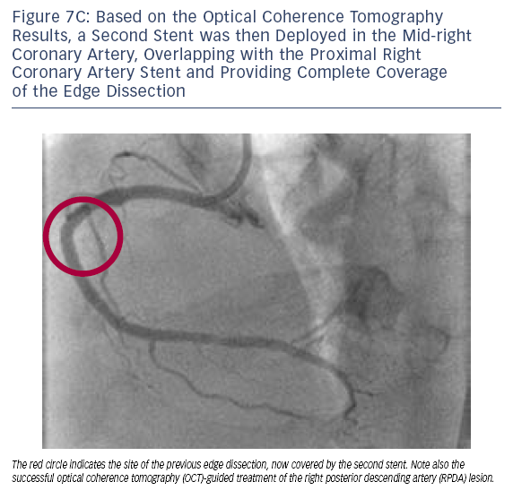
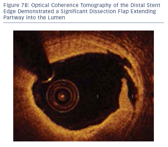
In this case, OCT was able to diagnose a significant distal edge dissection, guide the decision to implant a second stent and optimise PCI of the additional RPDA lesion.

Loading Simple Education